
ovarian cancer test
Surgical resection is the process by which the tumor is surgically removed, it is usually recommended when the cancer has not metastasized (or spread) beyond the lungs to other parts of the body.
There are several options for performing resection, which may include minimally invasive surgery, or full operation. What techniques are used will depend on various factors, particularly the size and stage of cancer and whether the tumor can be accessed by a surgeon.
thoracotomy the surgeon performs an incision through the chest wall and median sternotomy performed by entering the chest through the breastbone. Both of these methods are usually employed in lung cancer surgery, but they do not involve significant patient discomfort and extended hospital stays with more periods of recovery.
Alternative surgical procedures include anterior limited thoracotomy (ALT), which involves a small incision to allow entry, again through the front of the chest. It should be noted that the incision is much smaller than a standard thoracotomy or median sternotomija.Alternativa the anterior axillary thoracotomy (AAT), which involves a small incision on the chest front, but in the armpits and in the end, there is a postero-lateral thoracotomy (PLT) , which involves an incision on the back or side of the patient's trunk.
Even with better surgical techniques, the patient will experience considerable pain, if the operation involves opening the chest (sometimes known among doctors as "cracking the chest ").
43327, 43328 show the way for fundoplasty overhaul

only in open hernia repair codes to recognize the
Various ways your surgeon can perform the esophagogastric fundoplast - open or laparoscopic, across the chest and abdominal wall, with or minus hernia repair with mesh or minus. Here are the factors you will need to consider when attempting to select the appropriate code (s) from among nine choices in the CPT 2011th
Take a look at these four methods of advice for paraesophageal hiatalhernia repair and fundoplication coding for this year.
Tip 1: Know the pathophysiology
When a patient is said to have herniated, it typically means that part of the stomach is herniated through the opening in the diaphragm [Esophageal hiatus] in the chest and is usually associated with esophageal reflux disease.
Hernia repair typically involves surgery reducing the stomach back into the abdomen and stitching enlarged diaphragmatic hiatus. During the fundoplication procedure, say for example, Nissen, the surgeon wraps part fundus (top) of the stomach around the esophagus and sutured in place. This creates a 'valve', which allows food to go to the stomach from the esophagus, however, prevents reflux back into the esophagus.
Asbestos Cancer Exposure and Effects

asbestos cancer caused by asbestos exposure. Asbestos is a naturally occurring group of minerals that can be separated into thin threads. Asbestos fibers resist heat and fire, and for that reason, have been used in many products that need to be resistant to heat and fire, such as brake pads, gaskets and pumps. Unfortunately, however, asbestos-containing products, asbestos can cause cancer.
asbestos cancer: types
There are different types of asbestos cancer. They include lung cancer and mesothelioma. There are two types of lung cancer
Small cell lung cancer (SCLC) and
non-small cell lung cancer (NSCLC)
Small cell lung cancer is less common form of lung cancer and accounts for about 20% of cases of lung cancer. Small cell lung cancer occurs almost exclusively in smokers. It was more aggressive type of lung cancer and the time it is diagnosed, it usually has spread to many parts of the body.
non-small cell lung cancer is the most common type of lung cancer occur in approximately 80% of cases of lung cancer. Non-small cell lung cancer may have different causes, including exposure to asbestos.
Mesothelioma is a form of asbestos cancer. It affects the mesothelium, a membrane that covers the body of internal organa.Mesothelium produces fluid to allow the body slide against the adjacent struktura.Peritoneum the mesothelium that covers the organs in the abdominal šupljini.Pleura the mesothelium that covers the lungs and lines the chest wall koša.Perikard is the mesothelium that surrounds the heart.
The types of mesothelioma:
Pleural mesothelioma
The pleura is the lining of the bag prsima.Rak This lining is known as pleural mesothelioma. Pleural mesothelioma is the most common form of mesothelioma. Some symptoms of pleural mesothelioma are shortness of breath, chest pain and cough.
peritoneal mesothelioma
the peritoneum lining the abdominal lining abdomen.Rak is known as peritoneal mesothelioma. Peritoneal mesothelioma is less common than pleural mesothelioma. Symptoms of peritoneal mesothelioma include abdominal swelling, bowel obstruction, and nausea.
How can asbestos cause cancer, such as lung cancer and mesothelioma?
Origin of Connective Tissue Type Mesotheliomas from Multipotential Spindle Cells
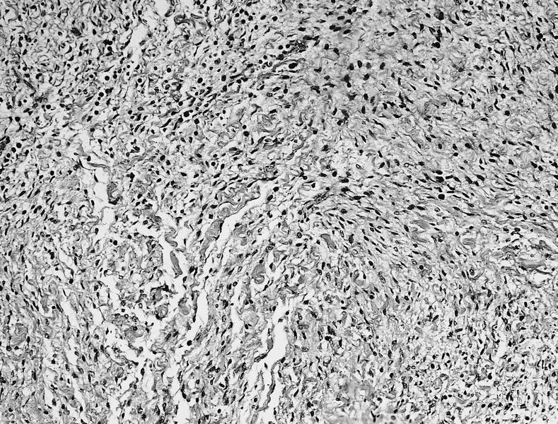
Another interesting study entitled "Pleural mesothelioma of connective tissue type, localized fibrous tumor of the pleura, and reactive hyperplasia submesothelial immunohistochemical comparison." Moutaiz by Al-Izzi, Nicola P. Thurlow, Professor Bryan Corrin -. Journal of Pathology, Volume 158, Issue 1, pages 41-44, May 1989 Here is an excerpt: "Abstract - Ten diffuse pleural mesotheliomas connective tissue type were compared with 14 examples of pleural granulation tissue and 7 localized fibrous tumors of the pleura, using immunohistochemistry to identify cytokeralins oflow and high molecular weight and vimentin. low-molecular-weight cytokeratin and vimentin are both delected in 8 of 10 mesotheliomas and 12 of 14 reactive iesions. cylokeratin high molecular weight is rarely detected or lesions. seven of localized fibrous tumors of the pleura are all positive and negative for virnentin for cytokeratins. These results support the origin of connective tissue type mesotheliomas multipotential submesothelial spindle cells and localized fibrous tumors of the pleura from any conventional fibroblasts or resting submesothelial stem cells. cytokeratin antibodies to help distinguish these two neoplasms, but does not provide help in difficult diagnostic problem of distinguishing mesotheliomas of the connective tissue type of pleural reaction is characterized by abundant granulation tissue ."
Another interesting study titled, "Final analysis of multi-center, double-blind, placebo-controlled, randomized phase II trial of gemcitabine / cisplatin (GC) plus bevacizumab (B) or placebo (P) in patients (pts ) with malignant mesothelioma (MM) "-
Another interesting study titled, "Final analysis of multi-center, double-blind, placebo-controlled, randomized phase II trial of gemcitabine / cisplatin (GC) plus bevacizumab (B) or placebo (P) in patients (pts ) with malignant mesothelioma (MM) "-
Saturday, August 27, 2011
Minimally Invasive Aortic Valve Replacement Through a Tiny Incision - See How it is Done

Da .. It is not rocket science! defective valve can now be repaired or replaced through a small incision in the chest. This procedure is known as minimally invasive aortic valve replacement. Many heart surgeons in the United States did not have time, ability or willingness to catch up with this revolutionary advancement in the field of heart surgery and will still offer the old-fashioned 12-14 "incision through the sternum (median sternotomy). It is often up to heart patients and their families to explore the demand for these advanced techniques of minimally invasive aortic valve replacement. If it is not available in your area, you are better off traveling to the center where it is routinely performed. This article will teach you everything you need to know to understand what surgery is available to you and to identify professional minimally invasive aortic valve heart surgeon.
This modern procedure is done safely and accurately through the 2 "incision between the ribs on the right side prsa.Rez is located just across the valve that needs replacing and is well known among surgeons as Minithoracotomy. Here is a list of the advantages of minimally invasive aortic valve replacement techniques of the old-fashioned breast bone splitting:
professional minimally invasive heart surgeon can perform all heart valve operations through minithoracotomy in over 90% of his patients. Always feel free to be very specific about what you want and ask many questions to the doctor. If you say that this procedure can be done in your case, ask why! Let me emphasize this point: the vast majority of patients who need aortic valve surgery are suitable candidates for minimally invasive aortic valve replacement. in expert hands there are no restrictions on age, overall condition of the patient or body weight. Insist on it. It's your body, your heart is, it's your life!
Symptoms, Diagnosis And Treatment Of Epithelioid Mesothelioma

sizcache = "0" sizset = "17">
Mesothelioma is a serious disease of cancer with low survival rates despite a variety of new treatment options that have helped to increase longevity. Statistics show that the average survival time after diagnosis is about one dana.Razdoblje can increase up to two years if the treatment is carried out aggressively. It was found that more than 95% of cases, mesothelioma is caused by exposure to asbestos.
Most people were previously aware of the dangers caused by exposure to asbestos as mesothelioma occurs after about 20-40 years after asbestos exposure. For this reason, this disease is also referred to as 'old people's disease', as people in 60 or 70 usually contract the disease. The higher forecast to help patients take better treatment, and in turn a better age.
epithelium is a tissue separating the different regions of the body. As in the case of epithelial cells in the skin are separates out the body from inside the body. This serves as protection for a diverse responsibilities, absorption, filtration, excretion, secretion, or sensory perception.
Causes and symptoms:
The most common cause of epithelioid mesothelioma is a place where fine particles and which are thrown into the air or by sawing or cutting. In epithelioid mesothelioma of the membrane lining the chest cavity, lungs, heart or abdominal cavity gets affected. In case of epithelioid mesothelioma patient survival time is about eight and a half months.
the rate at which a person is suffering as much as 50% to 70% of all cancer cells is the most common type. It was identified based on unique and organized cell structure which is tubular sample with a specific cell nucleus. Usually misdiagnoses diseases such cells in other cancers look very similar to the epithelioid cell adenocarcinoma is a type of cancer that is often confused with epithelioid mesothelioma.
is characterized by papillary or tubular growth, the most common subtype mestothelioma, there are many abnormal histological variants were found that include adenomatoid and deciduoid samples or tumor composed of small cells or mucin-positive cells, In addition to mesothelioma lepidic intrapulmonary growth.
Symptoms
Symptoms can occur after more than 40-50 years to be exposed to asbestos. This is most of the time mistaken for common asthma attacks, pneumonia or bronchitis because of symptoms of chest pain and breathlessness. Patients may have cough, weakness, fatigue, the inability to absorb nutrition causes weight loss. He also is a frequent gathering of fluid in the pleural space called the pleural effusion.The breath is caused when the tumor does not allow the lungs to expand and when spread on the results in severe chest pain.
Diagnosis
a thorough diagnosis is needed before any kind. As a latency period of this disease is the emergence of symptoms, early diagnosis becomes a difficult proposition. This is usually done only at an advanced stage leading to a survival rate of about 8-9 months.
To determine whether an individual has to undergo various diagnostic tests such as X-ray, MRI, CAT scan, Transbronchial biopsy, where the scope is passed through the trachea to the bronchi area of the lung, thoracotomy where the chest open and check the ribs for any cancer, thoracoscopy, where video cameras are used to check the ribs or Centesis where pleural, peritoneal, or pericardial fluid is removed.
Friday, August 26, 2011
Asbestos Cancer Exposure and Effects

asbestos cancer caused by asbestos exposure. Asbestos is a naturally occurring group of minerals that can be separated into thin threads. Asbestos fibers resist heat and fire, and for that reason, have been used in many products that need to be resistant to heat and fire, such as brake pads, gaskets and pumps. Unfortunately, however, asbestos-containing products, asbestos can cause cancer.
asbestos cancer: types
There are different types of asbestos cancer. They include lung cancer and mesothelioma. There are two types of lung cancer
Small cell lung cancer (SCLC) and
non-small cell lung cancer (NSCLC)
Small cell lung cancer is less common form of lung cancer and accounts for about 20% of cases of lung cancer. Small cell lung cancer occurs almost exclusively in smokers. It was more aggressive type of lung cancer and the time it is diagnosed, it usually has spread to many parts of the body.
non-small cell lung cancer is the most common type of lung cancer occur in approximately 80% of cases of lung cancer. Non-small cell lung cancer may have different causes, including exposure to asbestos.
Mesothelioma is a form of asbestos cancer. It affects the mesothelium, a membrane that covers the body of internal organa.Mesothelium produces fluid to allow the body slide against the adjacent struktura.Peritoneum the mesothelium that covers the organs in the abdominal šupljini.Pleura the mesothelium that covers the lungs and lines the chest wall koša.Perikard is the mesothelium that surrounds the heart.
The types of mesothelioma:
Pleural mesothelioma
The pleura is the lining of the bag prsima.Rak This lining is known as pleural mesothelioma. Pleural mesothelioma is the most common form of mesothelioma. Some symptoms of pleural mesothelioma are shortness of breath, chest pain and cough.
peritoneal mesothelioma
the peritoneum lining the abdominal lining abdomen.Rak is known as peritoneal mesothelioma. Peritoneal mesothelioma is less common than pleural mesothelioma. Symptoms of peritoneal mesothelioma include abdominal swelling, bowel obstruction, and nausea.
How can asbestos cause cancer, such as lung cancer and mesothelioma?
Lung Cancer Surgery Options

of lung cancer can be treated with various therapies that are often used in combination to provide the optimum outcome for the patient. Surgical resection is the process by which the tumor is surgically removed, it is usually recommended when the cancer has not metastasized (or spread) beyond the lungs to other parts of the body.
There are several options for performing resection, which may include minimally invasive surgery, or full operation. What techniques are used will depend on various factors, particularly the size and stage of cancer and whether the tumor can be accessed by a surgeon.
thoracotomy the surgeon performs an incision through the chest wall and median sternotomy performed by entering the chest through the breastbone. Both of these methods are usually employed in lung cancer surgery, but they do not involve significant patient discomfort and extended hospital stays with more periods of recovery.
Alternative surgical procedures include anterior limited thoracotomy (ALT), which involves a small incision to allow entry, again through the front of the chest. It should be noted that the incision is much smaller than a standard thoracotomy or median sternotomija.Alternativa the anterior axillary thoracotomy (AAT), which involves a small incision on the chest front, but in the armpits and in the end, there is a postero-lateral thoracotomy (PLT) , which involves an incision on the back or side of the patient's trunk.
Even with better surgical techniques, the patient will experience considerable pain, if the operation involves opening the chest (sometimes known among doctors as "cracking the chest ").
As a result of extended recovery time and patient discomfort, surgical techniques have been developed that do not involve full-blown operation -. These are the so-called minimally-invasive techniques
Video-assisted thoracoscopy (VAT) uses high-powered video cameras and hi-definition screen in combination with diagnostic scans such as CT or PET scans, to target tumors in the lung bolesnika.Rez should be much smaller and no need to open the chest, resulting in less discomfort for patients and greatly reduced the recovery period. Using the video display, the surgeon can resect tumors that were identified during the diagnosis and staging phases.
Some doctors caution the use of VAT, however, as the traditional thoracotomy can detect tumors and other cancers that metastasized were discovered in the initial scan and diagnosis. If you remain undetected cancer can return and the patient will become sick again and for that reason, the VAT is usually recommended for early stage (I and II), cancer, and not spread to other parts of the lungs and body.
It is normal for any surgical procedure to be followed by a phase of chemotherapy or radiation to ensure that the patient's cancer is completely removed or killed. Since treatment of income and in which phase the recommended treatment is determined by the type and stage of lung cancer the patient has.
Introduction to VATS Lobectomy Lung Surgery

There are more than 200,000 diagnosed cases of lung cancer (LC) each year in the United States. Although the incidence rate has been declining since 1991, the disease remains the leading cause of cancer-related deaths. When the condition is detected in stage 1 (ie, localized in the lungs), it can be treated successfully. By the time the cancerous cells have metastasized and spread to distant lymph nodes, it becomes harder to contain.
In the past, the disease is treated by thoracotomy (also known as open chest surgery). The patient's thoracic cavity access surgeon after a long incision is made in prsima.Prsne was cut and the ribs are spread to provide working space. Since the ribs have limited flexibility, the process sometimes resulted in fractures. To resolve this problem, thoracic surgeons began to remove parts of the patient's chest.
Today, minimally invasive techniques are often used to treat lung cancer. One of these techniques is a VATS lobectomy. This article will give an overview of the procedure and describe what to expect during recovery.
As the operation?
lobectomy is the surgical removal of the lobe (ie the portion of the lung). This is one of the most common procedure today for treatment of early LC. With VATS lobectomy, four incisions are made in the patient's chest. Three of the four sections are usually much less than one inča.Četvrti section can be measured to a few centimeters. No need to cut through the sternum and spread rebara.Cijeli procedure is carried out through these small incisions.
the surgeon will insert a thoracoscope, and several other surgical instruments through rezove.Thoracoscope is equipped with small lights and kamere.Fotoaparat transmits images back to the monitor uses the surgeon to see patients in the thoracic cavity.
The tumor is cut away from healthy tissue and extracted through one of the incisions. If the surgeon suspected cancerous cells have spread to nearby lymph nodes (ie the disease is in stage 2), some of them will be biopsied. This is done to confirm or disprove the spread of lung cancer to the lymph nodes.
Once the diseased tissue is removed, the surgeon will make sure that there are no signs of bleeding within the thoracic cavity. Four incisions are then closed and cleaned before the patient was transferred to a hospital recovery unit.
common questions about the work of
Before going through the screening process and go through the procedure, because patients often have many questions. For example, given the success rate for this type of lung cancer surgery and minimally invasive nature, some people wonder why they do not spend all bolnice.Odgovor is twofold. First, relatively few surgeons have mastered the techniques involved in VATS lobectomy. Second, some surgical centers lack the necessary equipment.
Another common area of confusion involves the patient's candidacy. Many people think this form of minimally invasive surgery of lung cancer can be performed on any person who is suffering from the disease. In reality, the procedure is generally reserved for those with stage 1 non-small cell LC.Tumor be measured at least three centimeters and remain localized in the lungs.
the recovery period after surgery is usually less than six tjedana.Pacijent will usually remain in hospital for two or three days after surgery. Then, he or she is released to complete their recovery at home. By the fourth week, most patients are able to return to their normal activity levels, although some require a full six weeks.
VATS lobectomy offers many advantages over traditional surgery, lung cancer was done through torakotomije.Vremena recovery is shorter, with less postoperative pain. In addition, the patient was able to return to normal life faster. If you are suffering from early stage lung cancer, ask your doctor whether you are a candidate for this approach.
Mesothelioma Surgery- What Are Your Options?
There are three main types of surgery used to treat mesothelioma.
Diagnosis-operation: This is used to confirm diagnosis and tumor location. It is usually noninvasive {it does not require cutting the patient's surgical}
B-curative surgery: This involves removing as much tumor as possible with the hope of treating the patient. Radiation therapy and chemotherapy and is often used in combination with this type of surgery.
C-Palliative surgery: This type of surgery offers only symptomatic relief. This involves the removal of cancer tissue, but does not offer a cure.
These are different types of surgical procedures for treatment:
1-Biopsy
This is a diagnostic form of surgery in which the suspected cancer tissue was partially removed and sent to a laboratory for research under a microscope to determine whether cancer cells. There are three types of biopsy procedures} cone biopsy is not generally relevant in the diagnosis of mesothelioma. b} Excisional biopsy involves removing as much as possible all the cancerous cells for testing. c} needle biopsy usually uses a long needle to remove a sample of cells from the area where cancer is suspected.
Thoracentesis 2-
Tuesday, August 23, 2011
43327, 43328 show the way for fundoplasty overhaul

only in open hernia repair codes to recognize the
Various ways your surgeon can perform the esophagogastric fundoplast - open or laparoscopic, across the chest and abdominal wall, with or minus hernia repair with mesh or minus. Here are the factors you will need to consider when attempting to select the appropriate code (s) from among nine choices in the CPT 2011th
Take a look at these four methods of advice for paraesophageal hiatalhernia repair and fundoplication coding for this year.
Tip 1: Know the pathophysiology
When a patient is said to have herniated, it typically means that part of the stomach is herniated through the opening in the diaphragm [Esophageal hiatus] in the chest and is usually associated with esophageal reflux disease.
Hernia repair typically involves surgery reducing the stomach back into the abdomen and stitching enlarged diaphragmatic hiatus. During the fundoplication procedure, say for example, Nissen, the surgeon wraps part fundus (top) of the stomach around the esophagus and sutured in place. This creates a 'valve', which allows food to go to the stomach from the esophagus, however, prevents reflux back into the esophagus.
Monday, August 22, 2011
43327, 43328 show the way for fundoplasty overhaul
only in open hernia repair codes to recognize the
Various ways your surgeon can perform the esophagogastric fundoplast - open or laparoscopic, across the chest and abdominal wall, with or minus hernia repair with mesh or minus. Here are the factors you will need to consider when attempting to select the appropriate code (s) from among nine choices in the CPT 2011th
Take a look at these four methods of advice for paraesophageal hiatalhernia repair and fundoplication coding for this year.
Tip 1: Know the pathophysiology
When a patient is said to have herniated, it typically means that part of the stomach is herniated through the opening in the diaphragm [Esophageal hiatus] in the chest and is usually associated with esophageal reflux disease.
Hernia repair typically involves surgery reducing the stomach back into the abdomen and stitching enlarged diaphragmatic hiatus. During the fundoplication procedure, say for example, Nissen, the surgeon wraps part fundus (top) of the stomach around the esophagus and sutured in place. This creates a 'valve', which allows food to go to the stomach from the esophagus, however, prevents reflux back into the esophagus.
Sunday, August 21, 2011
Minimally Invasive Aortic Valve Replacement Through a Tiny Incision - See How it is Done

Da .. It is not rocket science! defective valve can now be repaired or replaced through a small incision in the chest. This procedure is known as minimally invasive aortic valve replacement. Many heart surgeons in the United States did not have time, ability or willingness to catch up with this revolutionary advancement in the field of heart surgery and will still offer the old-fashioned 12-14 "incision through the sternum (median sternotomy). It is often up to heart patients and their families to explore the demand for these advanced techniques of minimally invasive aortic valve replacement. If it is not available in your region, you are better off traveling to the center where it is routinely performed. This article will teach you everything you need to know to understand what surgery is available for you and to identify professional minimally invasive aortic valve heart surgeon.
This modern procedure is done safely and accurately through the 2 "incision between the ribs on the right side prsa.Rez is located just across the valve that needs replacing and is well known among surgeons as Minithoracotomy. Here is a list of the advantages of minimally invasive aortic valve replacement techniques of the old-fashioned breast bone splitting:
professional minimally invasive heart surgeon can perform all heart valve operations through minithoracotomy in over 90% of his patients. Always feel free to be very specific about what you want and ask many questions to the doctor. If you say that this procedure can be done in your case, ask why! Let me emphasize this point: the vast majority of patients who need aortic valve surgery are suitable candidates for minimally invasive aortic valve replacement. in expert hands there are no restrictions on age, overall condition of the patient or body weight. Insist on it. It's your body, your heart is, it's your life!
Lung Cancer Treatment Options: Beating Lung Cancer

True to the greatest number of cases of cancer treatment depends on many factors. After lung cancer is set, the doctor and patient can discuss with each other now, the treatment options that will be needed. The patient should be well informed about the side effects and possible outcomes of certain procedures.
-
must all be addressed in order to avoid unpleasant. Other factors to take into consideration the general health status, health problems, medical treatment (eg chemotherapy) affect the characteristics of the tumor.
features lung cancer helps doctors to separate patients into two groups. Those with low risk of recurrence and those with high risk of recurrence of cancer
Mesothelioma Diagnosis

Mesothelioma is a technical term for cancer of the mesothelium.
It can be difficult to diagnose because many of the symptoms are similar to other conditions such as lung cancer.
your first visit to your doctor, he will ask about exposure to asbestos. Older houses are made of asbestos.
Although it is difficult to detect in its early stages, your doctor may perform several tests. Chest x-ray, CT and MRI scans allow the doctor to make a good diagnosis.
CT is used to create detailed images. MRI scan uses magnetism, radio waves and a computer, but does not use radiation to create a clear picture.
These tests help doctors differentiate between mesothelioma and other lung tumors. And they can also help determine where the tumor.
If needed, your doctor may need to draw the fluid to confirm the diagnosis. There are several ways to do it.
First, the doctor can make a small incision to place a flexible tube into the tumor, called thoracoscopy. Sometimes, however, more extensive surgical procedure May be desirable.
Second, thoracotomy can be done to open the chest to get a tissue sample, and to remove most of the visible tumor. Also, mediastinoscopy can be done to determine what a cancer.
Subsequently, samples of tissue under the microscope to determine whether it is mesothelioma cancer or other cancers.
I decided to write about mesothelioma because my dad was exposed to asbestos while working on the house. Now, he's breathing to be done.
Mesothelioma Research Limitations and Computed Tomography

Another interesting study titled "Impact of drug light interval on photodynamic therapy with meta-tetrahydroxyphenylchlorin in malignant mesothelioma" Hans-Beat Ris, Hans Jörg Altermatt, Bernhard Nachbur, J. Charles M. Stewart, Qiang. Wang, Chang Kee Lim, Raymond Bonnett, Ulrich Althaus - International Journal of Cancer Volume 53, Number 1, p. 141-146, 2 January 1993 Here is an excerpt: "Summary - The influence of time interval (TI) between the drug and laser activation on the selectivity meta-tetrahydroxy-phenylchlorin (mTHPC)-mediated photodynamic therapy (PDT) for tumor tissue was evaluated in BALB / c nude mice bearing human malignant mesothelioma xenografts. the following IP administration of 0.3 mg / kg mTHPC, light dose of 10 J / cm2 and 0.1 W/cm2 was delivered at 650 nm in the tumor and the same size area of the hind legs after the fourth I2, 24 and 36 hours, and 2.3, 4.5 and 6 days in groups of 6 animals (surface radiation ). Then, 72 hours after the delivery of light, depth of necrosis was measured in the tumor and the skin and underlying muscle hind legs. Photosensitized necrosis occurred in the normal tissue of the TI with 4 hr 3 days and tumor TI with I2 to 4 GB days. therapeutic ratio of mTHPC-PDT significantly varied with the time interval between the drug and the laser activated, and was the largest in the span of three days. mTHPC concentrations measured in 3 control unirradiated animals at all time points in normal tissues and tumor tissues, and it was found that same in both tissues. the tissue concentration of mTHPC was limited use in relation to the prediction of photosen-sitizing effects in tumor model ."
Another interesting study titled "Impact of drug light interval on photodynamic therapy with meta-tetrahydroxyphenylchlorin in malignant mesothelioma" Hans-Beat Ris, Hans Jörg Altermatt, Bernhard Nachbur, J. Charles M. Stewart, Qiang. Wang, Chang Kee Lim, Raymond Bonnett, Ulrich Althaus - International Journal of Cancer Volume 53, Number 1, p. 141-146, 2 January 1993 Here is an excerpt: "Summary - The influence of time interval (TI) between the drug and laser activation on the selectivity meta-tetrahydroxy-phenylchlorin (mTHPC)-mediated photodynamic therapy (PDT) for tumor tissue was evaluated in BALB / c nude mice bearing human malignant mesothelioma xenografts. the following IP administration of 0.3 mg / kg mTHPC, light dose of 10 J / cm2 and 0.1 W/cm2 was delivered at 650 nm in the tumor and the same size area of the hind legs after the fourth I2, 24 and 36 hours, and 2.3, 4.5 and 6 days in groups of 6 animals (surface radiation ). Then, 72 hours after the delivery of light, depth of necrosis was measured in the tumor and the skin and underlying muscle hind legs. Photosensitized necrosis occurred in the normal tissue of the TI with 4 hr 3 days and tumor TI with I2 to 4 GB days. therapeutic ratio of mTHPC-PDT significantly varied with the time interval between the drug and the laser activated, and was the largest in the span of three days. mTHPC concentrations measured in 3 control unirradiated animals at all time points in normal tissues and tumor tissues, and it was found that same in both tissues. the tissue concentration of mTHPC was limited use in relation to the prediction of photosen-sitizing effects in tumor model ."
Another interesting study titled "Malignant mesothelioma of the pleura:. Clinical aspects and symptomatic treatment," Law MR, Hodson ME, Turner-Warwick M. Here is an excerpt: "Abstract - series of 140 patients with malignant pleural mesothelioma reported clinical presentation was postponed in the case without large effusion, but the tumor is extensive. the presentation, they showed thoracoscopy, thoracotomy, or computed tomography in all patients investigated. thoracoscopy was a useful diagnostic alternative to thoracotomy. with disease progression, mesothelial expansion was more important than distant metastases, which are usually too small and they rarely produce symptoms. skin tumor deposits in areas of previous invasive procedures are not causing pain or other clinical problems, and we believe that diagnostic and therapeutic procedures should not be withheld to avoid them. in the management recurrent pleural effusion, intrapleural bleomycin, preceded by desire, and after vacuuming, a useful alternative to surgery, pneumothorax, spontaneous or iatrogenic, it decortication Adequate pain relief is difficult .. radiation therapy and nerve block procedures were not effective and the drugs are often necessary. "
We all owe debt of gratitude to these fine researchers. If you find any of these statements interesting, please read the study in its entirety.
Saturday, August 20, 2011
Lung Cancer Diagnosis - How This Works

What makes the diagnosis of lung cancer? doctor evaluates a person's medical history, smoking history, exposure to environmental and work substances, a family history of cancer, as well as physical examination and chest X-ray to find the cause of symptoms. Other tests may be performed as needed.
the patient's history - if the doctor suspects lung cancer, they will: explore the medical history, perform a thorough physical examination, in order to further specialized medical tests. As part of its history, the doctor will ask you: If you smoke or have smoked before, your interest and
Place of work: if you were exposed to hazardous substances at work or radiation. Do you have a family history of lung cancer
Diagnosis of lung cancer
Screening helps find cancer at an early stage when cure is a series of tests before a person shows any symptoms. Early detection of abnormal tissue or cancer confirms the favorable treatment of the cancer completely unlike the detection of symptoms when the cancer could spread.
There are several ways to diagnose if someone is in the early stages of cancer pluća.Fizički examination and medical history: physical examination, checking general signs of health or disease, such as disease and unusual lumps, bumps and everything else that makes atipične.Liječnik will also get a history of personal health habits, any past illnesses and treatments given to those diseases.
Laboratory tests procedures for testing of tissue samples, blood, urine and other substances in tijelu.Testovi will also help in diagnosing the disease, as well as assist in planning, management and control of it.
sputum test : This may show evidence of cancer cells in the lungs. To ensure more accurate diagnosis with a single sputum collection, sputum is usually collected for three days.
optic bronchoscopy : an overview using a small flexible lighted tube to pass into the channel of the nose, then the corresponding bronchi (airways) to cancer. If cancer is detected, then a small piece of cancer was removed for biopsy examination so that the exact type of cancer can be determined and appropriate treatment given.
Percutaneous needle biopsy : This exam involves inserting a thin needle through the skin and chest into the tumor. This is a test for tumors that are near the surface of the lungs and is often used in conjunction with a CAT scan helps in guiding the needle into the tumor.
excision or surgical removal of the : This process can lead to further diagnosis of suspected tumor through a small incision in prsima.Mali thin video camera is inserted into the chest to assist in removing small block of lung tissue by mechanical surgical stapling device or laser with the clinical procedure.
Mediastinoscopy : This test helps to assess how extensive the tumor is looking into the middle of the chest through a small incision just below the collar line. Samples were taken from the lymph nodes in the middle of the chest (mediastinum). The chances for surgical treatment of lung cancer is automatically eliminated, if the cancer has spread to lymph nodes.
Mediastinotomy : Unlike mediastinoscopy, chest cavity is cut open the sternum (breastbone) and / or ribs allows the surgeon to reach and test more lymph nodes by removing samples of mediastinal lymph nodes. It is a complex test, a patient must undergo general anesthesia.
Thoracentesis . A sample of fluid around the lungs take a needle to check for cancer cells
thoracotomy . To test for malignancy in the chest must be opened to the procedure performed in the hospital as one of the major operations
thoracoscopy . Procedure using a thin, lighted tube connected to video cameras to monitor and review the space between the lungs and chest
bone marrow biopsy with a needle sample of bone is removed usually measuring about 1 / 16 inches across and 1 inch long. This is often taken back with the hip bone. A microscopic sample is checked for cancer cells. This procedure is performed primarily to diagnose small cell lung cancer.
blood test : complete blood test checks for the exact number of different cell types that show whether you have anemia or other related problems. Blood tests show chemical abnormalities in the organs and other body parts. Blood tests are repeated regularly, especially if someone is undergoing chemotherapy. Chemotherapy drugs affect blood-forming bone marrow cells, and sometimes cause a lot of problematic side effects. If the cancer has spread to his liver and bones, it could cause certain chemical abnormalities in the blood and exacerbate the problems already suffered a patient.
Other tests and procedures for the detection of lung cancer include:
Chest x-ray : Chest x-rays make up about half of all x-rays obtained at bolnicama.X-rays are usually done in order to get an evaluation of your lungs, heart and koš.Prsima pelvic x-ray test is the first doctor to look for any tumor or of the lungs. If it is normal there is probably a big no lung cancer, but if anything suspicious is detected, the doctor will order additional tests. Pneumonia, heart failure, emphysema, other medical conditions, and lung cancer can be located with the chest x-ray.
CT scan or Computed Tomography also known as CT or CAT scan : This equipment has been available over-sectional images of organs and tissues in tijelu.MAČKA scanning particularly useful for diagnosing the tumor as which is far more detail than conventional chest x-ray. It shows the different types of body tissues, including lung, heart, bone, soft tissue, muscle and blood vessels at the same time.
Modern CT scan images of breast from different angles using a method called spiral (or helical) CT. With the help of computers, processes images to create sectional images or "slices" of the area causing the concern. Images can then be printed or viewed on a monitor. To obtain a better picture after the first set of scans are taken intravenously, the radio-contrast agent was administered through a review of structures within tijela.Drugi set of pictures then it takes so that they can be examined together.
information on the size, shape and position of the tumor provide CT. This helps detect any enlarged lymph nodes, which could contain cancer that has spread from the lungs. When searching for early lung cancer and to ensure patients receive the treatment they need as quickly as possible, CT is much more sensitive than ordinary routine chest x-ray. In the search for tumors in the adrenal glands, brain and other internal organs most commonly affected lung cancer spread to CT is also useful.
Magnetic Resonance Imaging (MRI) : MRI use radio waves and strong magnets instead of x-zraka.Energije released from the radio waves is absorbed and re-released in a pattern shaped by the type tissues and disease under investigation.
pattern of radio waves is given by the tissues and organs are highly detailed images of body parts using a very sophisticated computer. It can also produce slices parallel to the length of his body as a CT scanner produces cross-section slices of the body.
positron emission tomography (PET) : This scan uses glucose, which is a form of sugar that contains a radioactive atom. Large quantities of radioactive sugar is absorbed into cancer cells, and a special camera is then able to detect radioactivity.
to find out if someone is suffering from early stage lung cancer PET scan is a very useful test. Often used to detect if the cancer has spread to lymph nodes. Fri scans are valuable in determining whether the shadows on chest x-ray is cancer or not. Fri scans are also helpful when the doctor thinks the cancer has spread, but not sure where the spread can be.
Given that PET scans to scan your whole body around the place of several different x-rays. Bone scan: a radioactive substance (usually technetium diphosphonate) is injected into venu.Radioaktivne substance accumulates in areas of bone is suspected of having cancer metastasis (spread). Because small amounts of radioactivity used to not cause any long-term consequences.
Bone scan results should be read in conjunction with the results of other tests performed, and other bone diseases can also cause an abnormal scan results. Bone scans are usually performed on patients with small cell lung cancer, and also in non-small cell lung cancer patients when other test results or symptoms suggest the cancer has spread to the bones - the diagnosis of lung cancer
Thursday, August 18, 2011
Approaches to Minimally Invasive Mitral Valve Repair
When doctors diagnose problems with the mitral valve (MV), May they consider it necessary to solve them surgically before your heart is held fixed oštećenja.MV located between the left atrium and ventricle. Its mission is to facilitate the flow of blood from the atria (upper chamber) to the chamber (lower chamber), while preventing backflow. Unfortunately, there are several circumstances that prevent him performing his job effectively.
For example, one of his two leaflets can be loose or unsupported. Or, flyers can be elastic, causing them to flop (a condition known as pelvic organ prolapse). When these problems are present, the surgeon may decide that a candidate for minimally invasive mitral valve repair. We will explain several approaches to your surgeon can take to repair the MV without cutting completely through the sternum.
A partial upper sternotomy
A partial upper sternotomy
...To be fair, partial upper sternotomy is not technically considered "minimally invasive" because it requires cutting the sternum. But it is far less invasive than conventional open chest surgery. Often used in cases where the patient needs to have both her aortic and mitral valves repaired.
Right Mini thoracotomy
The right mini-thoracotomy, the sternum is cut at all. Instead of an incision made in the middle of the chest, an incision is made in your skin in the right prsima.Rez is usually three inches or less and allows the surgeon to insert the tools to approach the MV rebra.Potrebne repairs can be performed without the surgeon to directly access the heart.
robot-assisted mitral valve repair
This is a newer approach to minimally invasive mitral valve popravak.Kirurg will make a few incisions in the right chest. Two sections will be very small - two or three centimetra.Treći cut will be measured less than three inča.Kirurg will insert the robot arm through the small incisions. On the arms control computer, while watching the video monitor.Ruke sites are equipped with the necessary tools to slice through the pericardium and perform repairs.
approach, your surgeon decides that it will depend on the type of surgery you need, which valves are affected, how advanced the condition and the available tools. Minimally invasive mitral valve repair, although more widespread, it remains to be established in all hospitals. As always, consult with your doctor to discuss your surgical options.
Sunday, August 14, 2011
Scoliosis Surgery - Risks of Refusing Fusion Surgery for Adolescent Idiopathic Scoliosis Overstated

sizcache = "0" sizset = "56">
Severe scoliosis can present problems in cardiopulmonary function. The definition of severe scoliosis varies depending on sources. Most authors consider 60-degree standard for scoliosis is called severe. As for the crushing of internal organs, most of the current literature states that it is very rarely less than 1%, and only in cases of over 100 degrees. There are two major factors that go into deciding whether or not to take your child to undergo a spinal fusion operacije.Prvi the aesthetic look that most orthopedic surgeons say the number one concern of patients and their parents before learning potential cardiopulmonary učinak.Drugi the fear of health problem not only in the immediate future, but down the road.
This fear is probably based on a conversation with his orthopedic doctor regarding scoliosis and harmful impact on the cardiopulmonary system. Most often in my experience, in consultation with parents of children who are progressing scoliosis is that their understanding of "risk" is that if they do not have the surgery are putting your child at significant risk of health problems and potentially death. I think it is very important to discuss this mindset before entering into something that will change your life forever child.
There are several parameters that are often not discussed with the parents that would indicate a higher risk compared to almost no rizika.Istraživanja convincingly show that the increased risk of lung function loss resulting from structural thoracic scoliosis with Cobb measurements more than 60 degrees in the frontal plane and a significant loss of normal kyphosis in the sagital plane of 50% or more. According to the Lenke classification would be a subcategory of about 18% of patients with AIS to get a surgical threshold is 50 degrees.
curves with apexes below T9 generally do not have any restrictive lung problems, because there is no deformity of the chest, a much lower spine rigidity. Thoracic curves with apexes more than T7 have also been excluded. Primary structural thoracic scoliosis with normal to slightly reduced kyphosis are also questionable. So it would be very unlikely that we could say that "all" children with large curves will have any kind of damage to lung function loss.
Therefore, each child does not show a measurable decline in lung function should not be subjected to by medical necessity and justification should be thoroughly aware of the procedure for cosmetic appearance only of the process is not correcting the existing loss functions but the medical assumption that they are correcting a potential problem. So I should have my kidney removed because it looks abnormal on ultrasound and MRI, but all of my kidney tests are normal Hmmm.
other issues currently being debated is whether or not scoliosis surgery actually improves pulmonary function in adolescent idiopathic scoliosis. They agreed that it was undoubtedly a short-term effects are significantly reduced lung function, but long-term studies are certainly inconclusive.
conclusions. Pulmonary function after thoracotomy with ASA instruments showed a significant decline from 3 months postoperative PFT values, but returned to the preoperative absolute value of the basic 2-year follow-up visits. percent of predicted values returned to within 95% of the primary two years postoperatively. Scoliosis surgeons should be aware of these findings when deciding on the approach. Spine 2000; 25:2319-2325
Notice of lung function returned to preoperative levels after two years, but scoliosis surgery did not improve lung function.
conclusions - forced vital capacity was reduced to long track in adult patients with idiopathic scoliosis who undergo anterior spinal surgery. Decline in FVC is small and is unlikely to be of clinical significance in patients with reasonable lung function in whom surgery is planned for the prevention of progression of the curve and improve cosmetic appearance and pain. However, surgical intervention should be undertaken in an attempt to improve lung function (Thorax 1996, 51:534-536).
If the only reason to perform medical scoliosis fusion surgery for lung function, then the patient must have a measurable dysfunction at the time of surgery and increased lung function post surgery operations to be considered is the need for medical procedural point of view, no?
This is not a question of trust a doctor's opinion, but a message to all patients and parents to learn all the facts, studies, research, and get informed before agreeing to a highly invasive procedure that alter the body structurally and can not be undone.
Dr. Brian T. Dovorany
Common Questions About Minimally Invasive Mitral Valve Repair

Your mitral valve is located between the heart the left atrium and left ventricle. It regulates blood flow between the two chambers. As the electrical signals from the sinus node causes the atrium contracts, the valve opens to allow blood flow to the ventricle. Then it closes again, seal the opening and prevent backflow.
It may malfunction due to stenosis or regurgitation. Stenosis is characterized by narrowing of the valve opening, which restricts the blood from flowing through. Regurgitation is defined as a leak. In any case, the surgeon may recommend a minimally invasive mitral valve surgery (MIMVS) repair. Below, we answer four questions that patients frequently ask about work.
To be distinguished from open chest surgery?
Many years ago, the operation is accomplished by opening the patient prsima.Dugi cut (ie, 6 to 8 inches) is made and a surgical saw was used to share prsne.Pacijent the heart should be stopped during the procedure, especially the heart-lung machine was used for blood circulation.
minimally invasive approach can be performed without division of the chest or stopping the heart. Instead, several small incisions in the right flank pacijenta.Kirurg accessing the site through the ribs and mitral valve repair is performed through incisions.
What are the risks?
As with any type of chest surgery, the risk of infection and blood loss during MIMVS. However, these risks are considerably greater during and after open thoracotomy.
receives a better replacement?
Whenever possible, the surgeon will choose to repair the malfunctioning mitral valve, but it zamijeniti.Razlog is because the presence of foreign material (in this case, the artificial valve replacement) within your body sharply increases the risk of infection and clotting. This is why patients who receive replacement heart valves are usually required to anticoagulants for the rest of your life.
That said, there are circumstances in which the valve is damaged to the point needed to be replaced. Moreover, in some cases, the surgeon may lack the necessary skills to effectively perform mitral valve repair surgery.
What happens after surgery?
the recovery process for minimally invasive mitral valve repair is shorter and easier than recovering from open thoracotomy. Since the sternum is not divided access to the heart, there is less likelihood of infection and blood loss. Postoperative pain is reduced and you will be released from the hospital in less time. And finally, scarring is limited to small incisions between your ribs.
Fortunately, a minimally invasive mitral valve repair is used in many hospitals and surgical teams more than ever. Its growing importance in leading hospitals provide significant benefits for the patient with regard to this type of surgery.
Diseases of the Pleura III - Tumors of the Pleura and Pneumothorax
Cancers of the pleura
Primary tumor (mesothelioma) is a rare, and secondary tumors are common. Mesothelioma can be benign or malignant more often. Malignant pleural diseases more common in individuals chronically exposed to asbestos. Secondary tumors arise from cancer of the bronchus, stomach, liver and other facilities. Malignant lesions in the pleura lead to hemorrhagic pleural effusion.
pneumothorax
The presence of air in the pleural cavity is known as pneumothorax. Pneumothorax can be spontaneous, traumatic, and iatrogenic (artificially). Another classification is to divide them into the open, closed, and the error pneumothorax (tension pneumothorax). In an open penumothorax a free rental on the surface of the lungs through which air is obtained from the pleural cavity during inspiration and expiration. In a closed pneumothorax, pleura can not communicate with the outside. In tension pneumothorax, there is a mistake the slot on the surface of the lungs through which air enters the pleural cavity, but can not escape. As a result, tension pneumothorax develops, leading to respiratory and cardiac embarrassment.
of a spontaneous pneumothorax : The result of rupture of pulmonary lesions leading to leakage of air into the pleura. Subpleural blebs or bullae, pulmonary tuberculosis, congenital cystic disease of the bronchi, chronic bronchitis with emphysema, bronchial asthma, pneumoconiosis, and Staphylococcal pneumonia are common medical causes. Fracture of the ribs, thoracotomy, lung puncture on a needle biopsy, and bronchial rupture are common surgical causes. Sometimes a pneumothorax is produced artificially for diagnostic radiology in lung or as therapy to arrest massive nehoptysis.Pluća fails to hilium, when air enters the pleura and abolishes the negative pressure.
Clinical features : attack with unilateral pleuritis dispneja.Osjećaj pain and something gave way, was blamed by many, shortness of breath and an unproductive cough to develop soon. In tension pneumothorax respiratory embarrassment and cyanosis may be noted vidljivo.Zahvaćene hand and it does not move with breathing. Mid-line tructures is shifted to the opposite side. Percussion note hyper-resonant. Breath sounds are absent in many cases. If air enters the pleural cavity as in tension pneumothorax and open pneumothorax amphoric breath sounds čuti.Posebna percussion phenomenon "coin sound". The money is kept securely These front of the chest is struck with the second money. Auscultation on the back side of the chest reveals a metallic note. Adventitous sounds like the clicking sounds synchronous with the heart can be heard in the left-sided pneumothorax. Differential diagnosis includes pneumothorax other painful conditions associated with dyspnea, such as myocardial infarction and pulmonary infarction. Severe emphysema, large bullae, and diaphragmatic hernia can cause problems in diagnosis.
Radiological features : radiological features are diagnostic of a well-developed slučaj.Zahvaćene hypertranslucent by the due collection of free air in the pleural cavity and the absence of normal lung markings. Outside the borders collapsed lung is seen as a sharp margin on the background air. There is a shift of the trachea and mediastinum to the opposite side. Chest X should be in an upright posture, so that even a small collection of air will not be missed.
Complications : Although in most cases, spontaneous pneumothorax is a simple, serious complications can develop in some cases. These are:
1st Severe cardio-respiratory embarrassment due to compression of normal lung, which displaced the mediastinum in cases of tension pneumothorax
2nd air embolism
3rd surgical emphysema
4th infection of the pleural cavity resulting in the formation of hydro-or pyo-pneumothorax;
5th Penumothorax on the opposite side of the pre-existing lung disease and
6th failure of the spread collapsed lung.
HydroPneumothorax
When both air and liquid are present in the pleural cavity, it is known as hydropneumothorax. This is usually the result of rupture of pulmonary lesions, leaving the air, and exudates in the pleural cavity. In many cases, it is caused by tuberculosis. Other causes include lung abscess, bronchiectasis, bronchogenic carcinoma, and trauma to the chest. Some cases of pneumothorax is converted to hydro-pneumothorax, when the discharge develops as a result of infection.
The hydro-pneumothorax, a higher level of horizontal fluid can cause numbness to show what moves you pateint was made to adopt a different position (shifting dullness). On shaking gently until the patient auscultating the air-liquid interphase, succussion splash is heard. Chest readiograph reveals horizontal upper level of the liquid with the findings of pneumothorax above it.
Management of pneumothorax
Small closed pneumothorax, which did not seriously symptomatic may remain alone with bed rest and analgesics. Since the air will be absorbed within a few days. Tension pneumothorax can present as life-threatening emergency. If the tension is relieved to let the air May a patient die of cardio-respiratory failure.
Emergency Management : let the air by inserting a needle into the second intercostal SCAE 2-3cm beyond the lateral border of sternum and is connected to a rubber tube which was led under water, in order to prevent re-entry of air (underwater seal). This procedure can be initiated outside the hospital in time. Often, the needle has a tendency to block and intrapleural pressure goes up. Injury to the lung surface, bleeding into the pleura, infection and surgical emphysema, and other harmful side effects. When pneumothorax reaccumulates blockade pins, May he be replaced by wider tires catheter. After removing the patients in the hospital, the ideal procedure for the establishment of the pleura drainage hose connected to an underwater pečat.Cijev must be in place for several days or weeks. Antibiotics are given to prevent secondary infection of the pleura. It is obviously an underlying condition is treated with certain medications. The recovery of lung function is facilitated by starting physiotherapy at an early stage. If the lung does not expand with the removal of air, vacuum pleural cavity can help. Surgical repair of the pleural surface may be needed in intractable cases.
Why Is A Lung Surgery Necessary?

lung surgery is a type of medical procedure being performed on patients with severe lung problems. This is done especially when the mass is located in the lungs. Likewise, it is done in cases where the lung wall is weak or fluids are collected inside the lungs. In such cases, lung surgery is necessary to assess the level of problems and treatment issues. It will be cases where part of the lung must be removed, or sometimes, all it would have to be withdrawn. This will depend on the surgeon's recommendations, such as the process necessary for the patient.
Lung surgery is necessary to evaluate lung problems, even if the non-invasive imaging tests such as chest x-ray, CT and MRI had already detected the massive, weak lungs or fluid in the wall plućima.Razlog is that these test images have no way of knowing if the mass is located within the lung is a benign or malignant, but. Only surgery can detect or estimate the depth of the problem.
lung surgery procedure involves entering the chest in order to access the lungs. There are two types of lung surgery may be available danas.Postupak thoracoscopy or thoracotomy. Thoracoscopy is much easier and less invasive surgery procedures lung thoracotomy. At thoracoscopy, the surgeon uses a thin tube with a small camera and passes it through a small incision in the chest to see inside the lungs with a video monitor. There are times when thoracoscopy is done, the surgeon will insist to perform thoracotomy, too. Thoracotomy is the old way of doing lung operacije.Veliki incision was made on the chest when the surgery is done to see the lungs directly.
Mesothelioma and Pulmonary Decortication

Another interesting study titled "Pleurectomy for mesothelioma." Do Brancatisano RP, Joseph MG, McCaughan BC - Department of Surgery, Repatriation General Hospital Concord, NSW Med J Aust .. . 1st April 1991; 154 (7) :455-7, 460 Here is an excerpt: "Abstract - Objective: To evaluate the efficacy and safety of parietal pleurectomy in establishing a tissue diagnosis and control of pleural fluid accumulation in patients with pleural mesothelioma, and assess the success of this procedure caused mitigation DESIGN aND SETTING:. fifty consecutive patients with pleural mesothelioma who underwent thoracotomy for cardiac units in Concord and Royal Prince Alfred Hospital were reviewed retrospectively male:. female ratio was 04:01 and mean age was 63 years in only 11 of 50 patients had tissue diagnosis of mesothelioma was before the surgery INTERVENTIONS: .. the thoracotomy, subtotal parietal pleurectomy was performed in 45 of 50 patients in two patients. biopsy only was performed, and three patients were treated by chemical pleurodesis only as pleurectomy is not technically possible . pulmonary decortication was required in 28 patients to allow full expansion of the underlying lung for effective pleurodesis.
RESULTS: There was one postoperative smrt.Morbiditeta was 16%. Without a patient who died in the postoperative period, median survival was 16 months, ranging from three to 54 months, with 21% of patients surviving for more than two years. Only one patient developed a reaccumulation of pleural fluid.
Conclusion: Pleurectomy with decortication if necessary, and provide tissue diagnosis and effective control of pleural fluid accumulation and thus a great reduction in patients with pleural mesothelioma. We advocate early thoracotomy in these patients ."
Thursday, August 11, 2011
Is Asbestos Killing Your Family?

sizcache = "0" sizset = "43">
Mesothelioma is a cancer that attacks the lining of the lungs and has a direct relationship with people who came into direct contact with asbestos.
mesothelioma is also known as asbestos cancer, strikes many, many thousands of people worldwide every year.
More than 3,000 people are diagnosed in the United States each year.
Once a person is diagnosed with mesothelioma are usually dead within two years.
people who are most likely to contract mesothelioma are usually workers who worked in factories, in mines, in manufacturing plants or are constantly in contact with asbestos or asbestos dust.
Mesothelioma symptoms include shortness of breath, cough and painful recurring chest and abdomen. Sometimes the patient feels my hard stabbing pain in the chest or abdomen.
Mesothelioma Symptoms usually surface only 35-50 years after asbestos contact. However, if an individual displays symptoms of mesothelioma they should consult a doctor immediately.
mesothelioma is usually diagnosed by chest X-ray, complete blood count, biopsy, or thoracotomy.
There are several different treatment options for people suffering from mesothelioma.
The most common form of treatment is surgery, where doctors will try to remove as much cancer as possible.
Palliative surgery is used when the disease is more advanced stage.It is used to control symptoms or experiences where the accumulation of fluid in the lungs or lung tumor pressing against the wall.
The radiation used to kill cancer calls and shrink tumors, works by firing high-energy X-rays at the tumor.
Radiation therapy has few side effects such as vomiting, nausea and general fatigue.
Chemotherapy is another treatment for mesothelioma cancer. Chemotherapy does not show a good success rate when it comes to treating cancer mesothelioma, turnout was about 15 %.
It is important to come to term with his cancer, so will make your life more comfortable so you do not live in anger, fear or sadness.
There are many groups offering assistance to people living with mesothelioma cancer, ask your doctor first.
Anthony Gregory helps promote the following sites:
Statistic
